Keratoconus
Progressive thinning of the cornea into a forward-bulging cone shape.
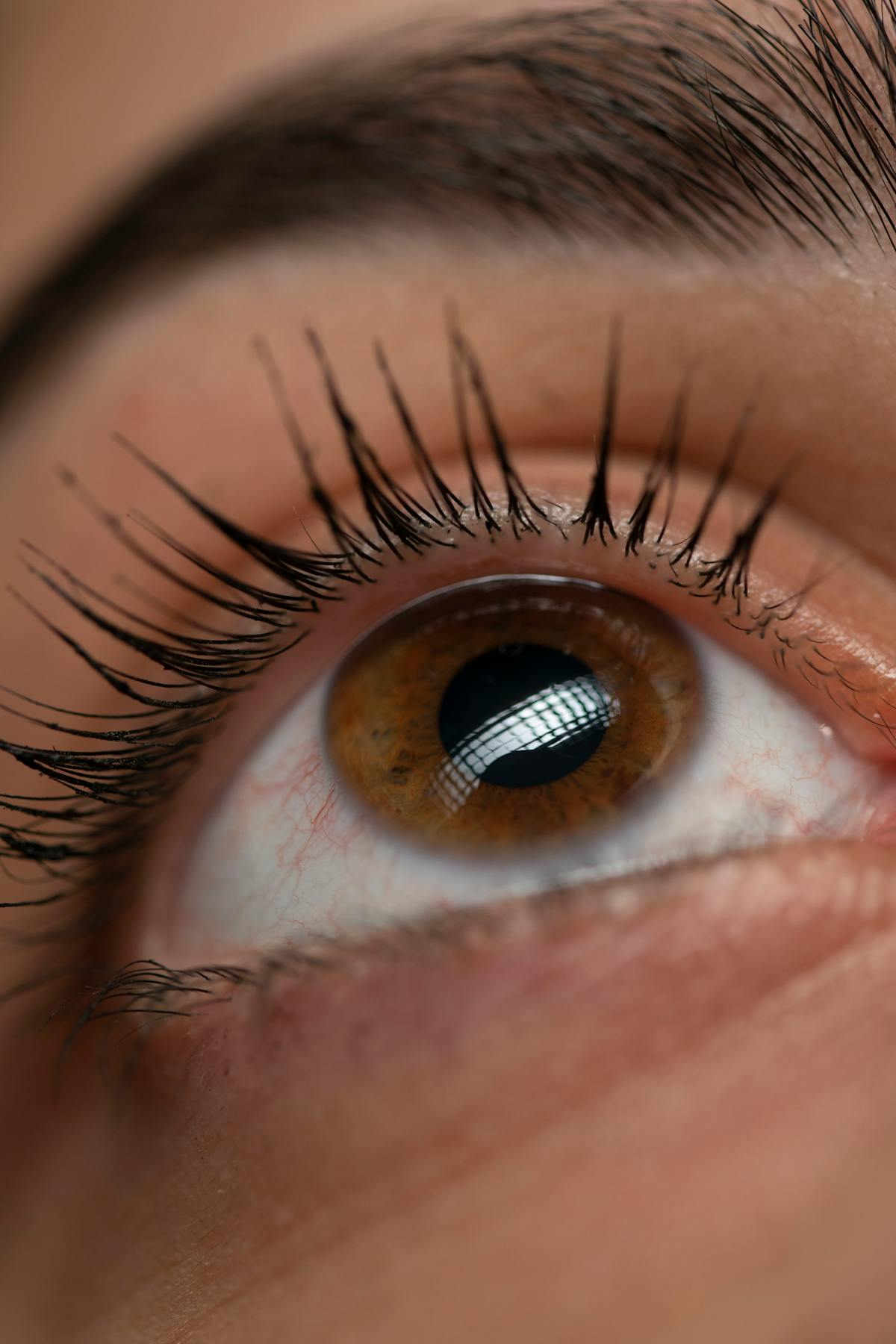
The cornea is the eye's front window — a curved, transparent dome that does most of the work of focusing light. In keratoconus, that dome thins and starts to bulge forward into a cone. Vision becomes irregularly distorted in ways glasses can't fully correct, often starting in the late teens.
Why early diagnosis matters.
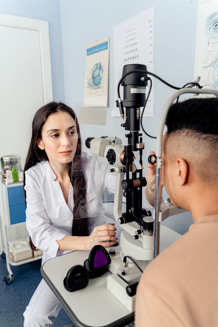
Untreated, keratoconus progresses for years and used to end, for some patients, in a corneal transplant. We now have a simple, in-office procedure — corneal cross-linking — that strengthens the collagen of the cornea and stops the progression in most patients, if we catch it early. Topographic imaging is what makes that catch possible.
How we treat it.
Early disease is corrected with rigid or specialised soft contact lenses that smooth out the irregular surface. Cross-linking halts progression. Where the cone is too advanced, intracorneal ring segments can flatten it; transplant is now a last resort, and rare.